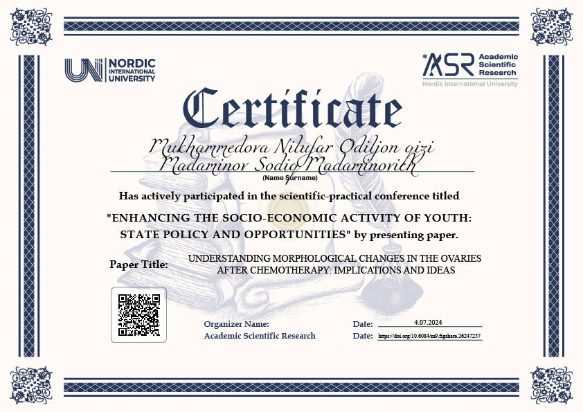
Mukhammedova Nilufar Odiljon qizi1,
Madaminov Sodiq Madaminovich2


Doi: https://doi.org/10.6084/m9.figshare.26247257
Figshare community: https://figshare.com/projects/_YOSHLARNING_IJTIMOIY-IQTISODIY_FAOLLIGINI_OSHIRISH_DAVLAT_SIYOSATI_VA_IMKONIYATLAR_/210568
Zenodo community: https://zenodo.org/records/12722584
Nordic_press journal: https://research.nordicuniversity.org/index.php/nordic/issue/view/5
ANTIPLAGIAT NATIJASINI YUKLAB OLISH
MAQOLANI YUKLAB OLISH
SERTIFIKATNI YUKLAB OLISH
REVIEW:
"Understanding Morphological Changes in the Ovaries After Chemotherapy: Implications and Ideas"
The article "Understanding Morphological Changes in the Ovaries After Chemotherapy: Implications and Ideas" provides a comprehensive analysis of the effects of chemotherapy on ovarian morphology, highlighting the significant impact on fertility and overall reproductive health. Authored by Nilufar Odiljon qizi Mukhammedova and Sodiq Madaminovich Madaminov from the Fergana Medical Institute of Public Health, the paper presents a systematic review of existing literature, clinical observations, and experimental data to elucidate the underlying mechanisms and broader implications of chemotherapy-induced ovarian changes.
Summary:
The article begins by emphasizing the crucial role of ovaries in female reproductive health and the profound structural alterations they undergo following chemotherapy. The authors meticulously discuss the cytotoxic effects of chemotherapy agents on ovarian cells, particularly primordial and growing follicles, leading to diminished ovarian reserve over time. This depletion is closely associated with fertility issues and hormonal imbalances.
Methodology:
The study employs a robust methodology combining a systematic literature review, clinical observations, and experimental data analysis. This multifaceted approach allows the authors to provide a detailed understanding of the morphological changes in ovarian tissues due to chemotherapy. The use of high-resolution imaging techniques, such as transvaginal ultrasound, Doppler studies, and MRI, alongside biomarkers like AMH and inhibin B levels, offers a comprehensive evaluation of ovarian reserve and tissue architecture post-chemotherapy.
Key Findings:
Mechanisms of Chemotherapy on Ovarian Morphology:
- Chemotherapy agents induce apoptotic cell death in ovarian cells, particularly affecting primordial and growing follicles, thus reducing ovarian reserve.
Diagnostic Methods for Assessing Morphological Changes:
- Advanced imaging techniques and biomarkers are crucial in assessing the extent of ovarian damage and predicting fertility outcomes.
Prognostic Value of Morphological Changes:
- The extent of follicular depletion and ovarian fibrosis serves as a predictor for decreased ovarian reserve and increased risks of premature ovarian insufficiency.
Clinical Implications:
The discussion highlights the clinical relevance of understanding chemotherapy-induced ovarian changes for patient counseling and fertility preservation strategies. The authors suggest incorporating imaging and biomarker assessments into clinical practice to tailor personalized treatment plans and mitigate ovarian damage. They also underscore the need for interdisciplinary collaboration among oncologists, reproductive endocrinologists, and fertility specialists.
Future Directions:
The article concludes by suggesting avenues for future research, including advancements in imaging technology and exploration of novel biomarkers to enhance the precision of ovarian assessments and identify new therapeutic targets for mitigating ovarian injury.
Conclusion:
Overall, this article makes a significant contribution to the field of oncofertility by providing a thorough understanding of the morphological changes in the ovaries post-chemotherapy and their implications for women's health. The comprehensive analysis and practical recommendations offered by the authors are invaluable for clinicians and researchers alike, aiming to improve patient care and long-term reproductive outcomes.


